Landmarks In The Evolution Journey Of ECG Machine
Landmarks In The Evolution Journey Of ECG Machine
Electrocardiography is an essential part of the clinical investigation for patients presenting with cardiac complaints. Specifically, it plays an important role as a non-invasive, cost-effective tool to evaluate arrhythmias and ischemic heart diseases.
Electrocardiography is the process of producing an electrocardiogram (ECG or EKG), a recording of the heart’s electrical activity through repeated cardiac cycles. It is an electrogram of the heart which is a graph of voltage versus time of the electrical activity of the heart using electrodes placed on the skin. These electrodes detect the small electrical changes that are a consequence of cardiac muscle depolarization followed by re-polarization during each cardiac cycle (heartbeat). Changes in the graph compared with the normal ECG pattern are indicators of abnormalities, including, Cardiac rhythm disturbances, ventricular tachycardia, inadequate coronary artery blood flow (such as myocardial ischemia) and myocardial infarction.
ECGs are standard equipment in clinics, hospitals, and ambulances across the world which is reliable and painless way to quickly detect heart problems. Though healthcare sector is using ECG very commonly, its evolution journey from the concept to easy to use handy machine is very interesting and challenging also. It is the journey of decades which includes tremendous efforts of different scientists around the globe.
In the era of rapid technology, some personal devices, such as smartwatches and fitness trackers, offer ECG monitoring.
Why have ECGs become so standardized—influencing both cardiology and the overall field of medicine so significantly? Let’s retrospect the journey of evolution of the ECG, starting with its history that stretches back to the 18th century.
Timeline of clinically pertinent landmarks in the development of EKG (ECG)

- In the 1786, Dr. Luigi Galvani, an Italian physician and physicist at the University of Bologna, first noted that electrical current could be recorded from skeletal muscles. He recorded electrical activity from dissected muscles.
- In 1842, Dr. Carlo Matteucci, a professor of physics at the University of Pisa, demonstrated that electrical current accompanies every heart beat in a frog.
- In 1872, Alexander Muirhead is reported to have attached wires to the wrist of a patient with fever to obtain an electronic record of their heartbeat.
- In 1882, John Burdon-Sanderson working with frogs, was the first to appreciate that the interval between variations in potential was not electrically quiescent and coined the term “isoelectric interval” for this period.
Considering above findings of electrical activities within heart and through muscles, In 1887, Augustus Waller, a British physiologist of St Mary’s Medical School in London, published the first human electrocardiogram using a capillary electrometer and electrodes placed on the chest and back of a human. He demonstrated that electrical activity preceded ventricular contraction. It is the first time that cardiac electrical activity had been detected and recorded.
Augustus Waller invented an ECG machine consisting of a Lippmann capillary electrometer fixed to a projector. The trace from the heartbeat was projected onto a photographic plate that was itself fixed to a toy train. This allowed a heartbeat to be recorded in real time.
A few years later, physiologist and Nobel Prize winner Willem Einthoven improved on the electrometer, coining the term “electrocardiogram “and created a formula that made it possible to describe name and distinguish between cardiac deflections called the P wave, the QRS complex, and the T wave (PQRST).
The invention of the electrocardiograph by Dutch physiologist Willem Einthoven in 1902 gave physicians a powerful tool to help them diagnose various forms of heart disease, especially arrhythmias and acute myocardial infarction. In 1924, Einthoven was awarded the Nobel Prize in Medicine for his pioneering work in developing the ECG.
The discovery of x-rays in 1895 and the invention of the electrocardiograph 7 years later inaugurated a new era in which various machines and technical procedures gradually replaced the physician’s unaided senses and the stethoscope as the primary tools of cardiac diagnosis. These sophisticated new approaches provided objective information about the structure and function of the heart in health and disease.
In 1942, Emanuel Goldberger increases the voltage of Wilson’s unipolar leads by 50% and creates the augmented limb leads aVR, aVL and aVF. By adding 3 more leads for better accuracy, the final 12-lead electrocardiogram that is used today was formed.
In the late 1940s, Rune Elmqvist invented an inkjet printer involving thin jets of ink deflected by electrical potentials from the heart. It opened the door of direct recording of ECG on paper. The device, called the ‘ Mingograf’ was sold by Siemens Elema until the 1990s.
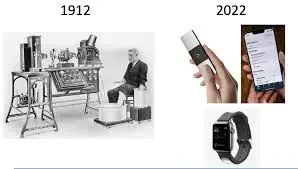
Willem Einthove is considered the founder and father of modern ECG. The earliest models purportedly weighed more than 600 pounds and required a five-person operator team. After that designs were changes according to parallel new discoveries of material, techniques etc.
The physiology, Patho physiologies and associated interpretation from graphs of cardiac activities from different limb had advanced in significant ways. From a clinical utility standpoint, the ECG has advanced over the period of time, from mid-century descriptions of tachycardia to the 1980s era of defining QRS with parameters that can be predictive of heart failure. Over time, ECG interpretations have also evolved to identify and inform diagnoses across cardiology including Coronary disease, coronary occlusion, cardiomyopathy, pericarditis or myocarditis, previous heart attacks and cardiac arrest, imbalances in electrolytes that control heart activity.
First ever electrocardiography (ECG) machine was used in K.E.M. Hospital, Mumbai in late 1940s. Two such mammoth size ECG machines are still available as antique specimen.
Today’s versions of machine is having typically around 8 pounds weight needing just one operator. The clinical accessories that accompany an ECG unit have evolved as well. Cables, lead wires, and thermal papers have all become more durable, reliable, and intuitive for users as the years progress.
In the first half of the 20th century, a number of innovative individuals set in motion a fascinating sequence of discoveries and inventions that led to the 12-lead compact electrocardiogram, as we know it now.
Here are some interesting facts related with the process of development of ECG machine
– Frogs played an important role.
Scientists measured electrical activity in frogs, and these creatures aided in many discoveries that eventually led to today’s ECG machines.
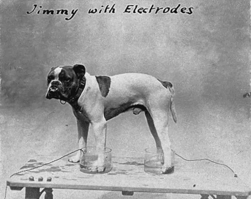
– Genius Dog
Augustus Desiré Waller was a British physiologist who used a capillary electrometer to record the first human electrocardiogram. His dog Jimmy often helped him demonstrate the electrogram by placing his paws in glass jars of saline.
– The first ECG machine weighed 600 pounds
Einthoven invented the first ECG machine In 1903. It was a large contraption that weighed 600 pounds. Today the machine is just a few pounds and even available in some smart watches.

– ECG machines used to be powered by car batteries
In 1928, Frank Sanborn introduced a more practical version of the EKG ( ECG ) machine. It was a table-top model that weighed 50 pounds and was powered by a 6-volt automobile battery.
Technology has become an integral part of our live in this era. Rapid advancement in medical devices for diagnostic, treatment, surgery purposes are helping healthcare sector very effectively.
Development of ECG is definitely great and inspiring milestone in the history of medical science which serves as pioneer work in advancement of many biomedical devices.
For regular updates, like and follow: